You perform an LP. Initial CSF results are notable for WBC 4 (65% PMNs), RBC 5, protein 95, glucose 40. A few hours later, HSV PCR comes back negative.
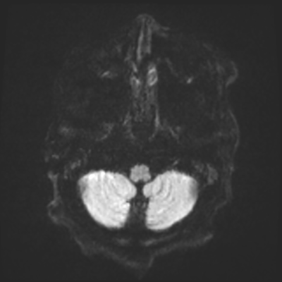
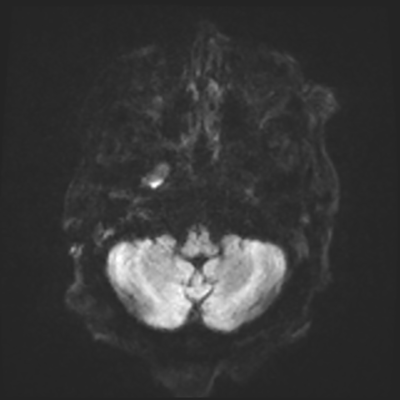
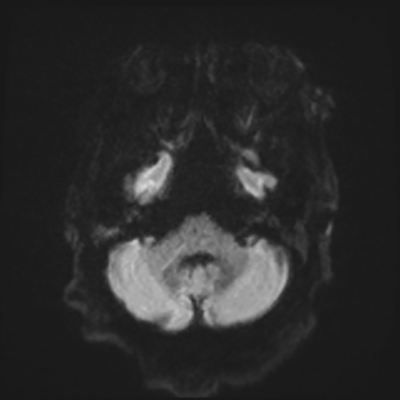
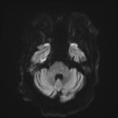
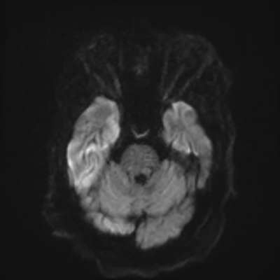
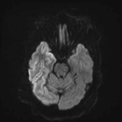
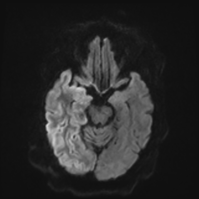
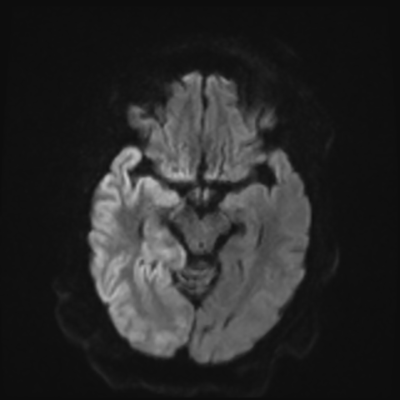
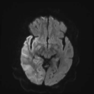
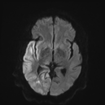
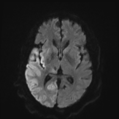
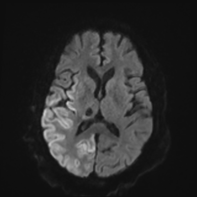
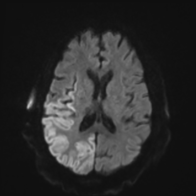
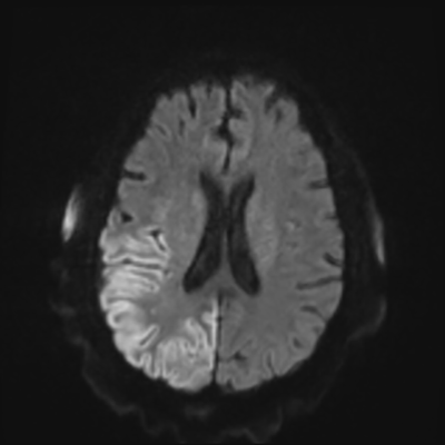
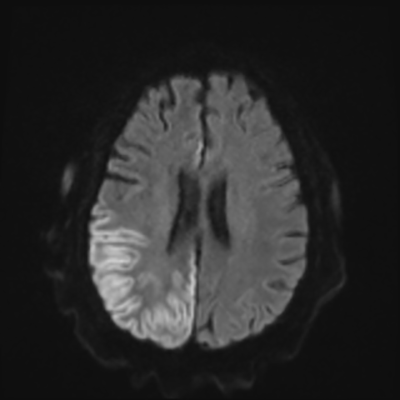
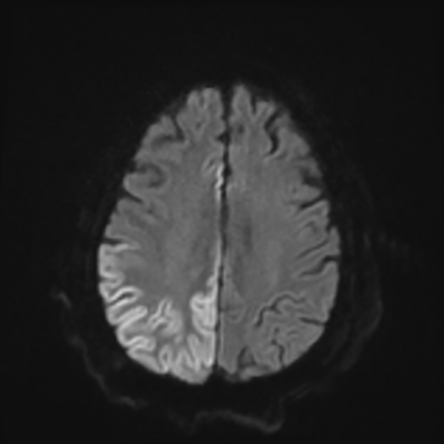
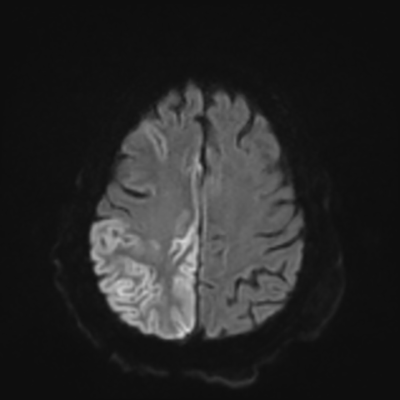
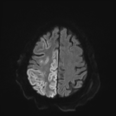
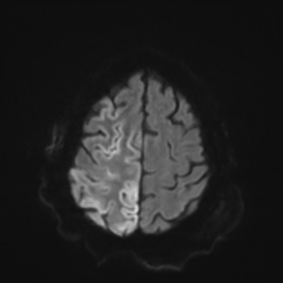
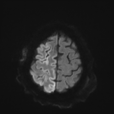
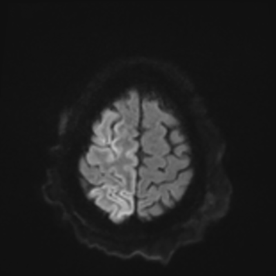
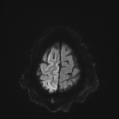
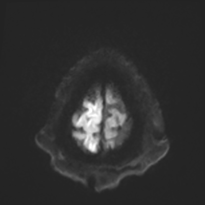
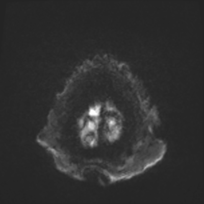
You come back the next day and see how things are going. The patient's electrographic record has shown a moderate encephalopathy (diffuse slowing, disorganization) with frequent right posterior quadrant delta slowing. Based on this result, and an adequately improved clinical examination, you give the okay to proceed with obtaining an MRI of his brain. This is shown below.
 1/24
1/24
 2/24
2/24
 3/24
3/24
 4/24
4/24
 5/24
5/24
 6/24
6/24
 7/24
7/24
 8/24
8/24
 9/24
9/24
 10/24
10/24
 11/24
11/24
 12/24
12/24
 13/24
13/24
 14/24
14/24
 15/24
15/24
 16/24
16/24
 17/24
17/24
 18/24
18/24
 19/24
19/24
 20/24
20/24
 21/24
21/24
 22/24
22/24
 23/24
23/24
 24/24
24/24
It's now day 2, and his CSF cultures remain no growth to date. He remains on cEEG, and the epilepsy fellow pages you to tell you that the patient is having 1Hz LPDs, maximal at F8 (do you want to disconnect?).
Copyright © 2024-2026 Andrew M. Nguyen, M.D. For educational use only.
This work is licensed under
CC BY-NC-SA 4.0.![]()
![]()
![]()
![]()